Meniscus Injuries  – Knee Pain – Physiotherapy in Tralee

Cartilage within the knee joint provides cushioning between the bones at this joint. This protects them from the stresses of walking, jumping, running etc. There is articular cartilage which is the smooth, white tissue that covers the ends of bones(Femur, tibia) where they come together to form the knee joint. Healthy cartilage in our joints makes it easier to move. It allows the bones to glide over each other with very little friction. Articular meniscus injuriescartilage can be damaged by injury or normal wear and tear.
Within the knee you also have fibrocartilage in the form of the medial and lateral meniscii. These are two thick wedge-shaped pads of cartilage attached to top of the tibia (tibial plateau) and under the femur bone. They  allow the femur to glide when the knee joint moves. Each meniscus is curved in a C-shape, with the front part of the cartilage called the anterior horn and the back part called the posterior horn. Meniscal tears are usually described by where they are located anatomically in the C shape and by their appearance (for example, “bucket handle” tear, longitudinal, parrot beak, and transverse).

Blood supply

Because the blood supply is different to each part of the meniscus. Knowing where the tear is located may help decide how easily an injury might heal (with or without surgery). The better the blood supply, the better the potential for recovery. The outside rim of cartilage has better blood supply than the central part of the “C.” Blood supply to knee cartilage also decreases with age, with up to 20% of normal blood supply  lost by age 40.

What causes a meniscus to tear?

A forceful twist or sudden stop can cause the end of the femur to grind into the top of the tibia, pinching and potentially tearing the cartilage of the meniscus. These meniscus injuries can also occur with deep squatting or kneeling, especially when lifting a heavy weight. Meniscus tear injuries often occur during athletic activities, especially in contact sports like football and hockey. Motions that require pivoting and sudden stops, in sports like tennis, basketball, and golf, can also cause meniscus damage.

Increasing risk with age

The risk of developing a torn meniscus increases with age. This is  because cartilage begins to gradually wear out, losing its blood supply and its resilience. Being overweight also puts more stress on the meniscii. This means that routine daily activities like walking and climbing stairs increase the potential for wear, degeneration, and tearing. It is estimated that six out of 10 patients older than 65 years have a degenerative meniscus tear. Many of these tears may never cause problems.

Some of the fibers of the cartilage are interconnected with those of the ligaments that surround the knee. Thus, meniscus injuries may also be associated with tears of the collateral and cruciate ligaments, depending upon the mechanism of injury.

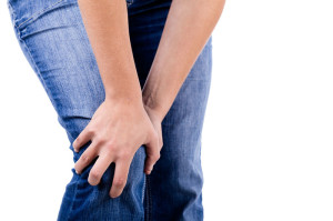
Symptoms of a meniscus injury can include some or all of the following:

  • Pain with running or walking longer distances
  • Intermittent swelling of the knee joint: Many times, the knee with a torn meniscus feels “tight.”
  • Popping, especially when climbing up or down stairs
  • Giving way or buckling (the sensation that the knee is unstable and the feeling that the knee will give way): Less commonly, the knee actually will give way and cause the patient to fall.
  • Locking (a mechanical block where the knee cannot be fully extended or straightened): This occurs when a piece of torn meniscus folds on itself and blocks full range of motion of the knee joint. The knee gets “stuck,” usually flexed between 15 and 30 degrees and cannot bend or straighten from that position.  

Diagnosis

The diagnosis of a knee injury begins with a history of the injury etc. and physical examination. There have been many tests described to assess the internal structures of the knee. The McMurray test is one long used orthopedic test . The health-care professional flexes the knee and rotates the tibia while feeling along the joint. The test is positive for a potential tear if a click is felt or noticeable pain is felt while circumducting the knee in full flexion.

Physiotherapy in Tralee – referral for MRI

(MRI) is the test of choice to confirm the diagnosis of a torn meniscus. It also allows a radiographer to visualize the inner structures of the knee. These structure include; the cartilage and ligaments, the surface of the bones, and the muscles and tendons that surround the knee joint. Plain X-rays cannot be used to identify meniscal tears but may be helpful in looking for bony changes, including fractures, arthritis, and loose bony fragments within the joint. In older patients, X-rays may be taken of both knees while the patient is standing. This allows the joint spaces to be compared to assess the degree of cartilage wear. Cartilage takes up space within the joint and if the joint space is narrowed, it may be an indicator that there is less cartilage present, likely from degenerative disease.

Treatment of Meniscal Tears

Sometimes conservative measures such as physical therapy, NSAIDs and rest can be enough to settle the condition. When conservative measures are ineffective the next step may be surgery to repair or remove the damaged cartilage.


For physiotherapy in Tralee phone 086-7700191, click here for website homepage
”Shin Splints” – Which Type Have You ?

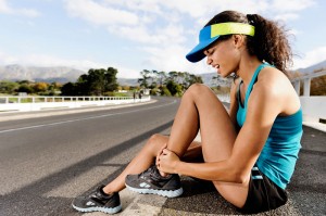
shin splints”Shin splints” is a catch-all term for shin pain either on the front outside part of the lower leg (anterior shin splints) or on the inside of the lower leg (medial shin splints). It is the curse of many athletes including runners, tennis players, dancers etc. Frequently the condition plagues novice runners who do not build their mileage gradually enough. It also affects seasoned runners who abruptly change their workout regimen, suddenly adding too much mileage, or switching from running on the flat to hills. The term mainly refers mainly to the following three conditions 1. mini stress fractures within the tibia bone, 2. chronic exertional compartment syndrome, 3. medial tibial stress syndrome.  It is important to differentiate between the three for treatment.


1. Real ”shin splints”

Real ”shin splints” are mini stress fractures (splint-ers) within the tibia bone. With this condition pain is gradual in onset, getting worse with activity, and there is usually a history of an increase in training intensity. Pain may occur with walking, at rest, or even at night in bed. Treatment for this condition involves rest for about eight weeks from running to allow the little stress fractures to heal. You should be able to keep up fitness levels by cycling, swimming etc., as these exercises are low impact. You may need to look at lower limb biomechanics, running style, training practices etc. to prevent recurrence of the shin splints .


2. Shin splints – Chronic exertional compartment syndrome

Chronic exertional compartment syndrome is defined as increased pressure within a closed fibro-osseous space(like the space the tibia and fibula), causing reduced blood flow and tissue perfusion(perfusion is the process of a body delivering blood to a capillary bed in its biological tissue), which subsequently leads to ischemic pain(pain due to restriction of blood supply, and thus oxygen and nutrients to tissue) and possible permanent damage to tissues of the compartment. The syndrome is frequently bilateral (both legs). Typical features of the condition are absence of pain at rest, with increasingly achy pain and a sensation of tightness in the shins upon exertion. Symptoms usually resolve or significantly dissipate within several minutes of resting. Anyone can develop the condition, but it is more common in athletes who participate in activities that involve repetitive impact, such as running. Sometimes Chronic exertional compartment syndrome may respond to deep tissue work and myofascial release of the structures involved. Changing your chosen activity to one involving less impact may also help. Surgery may be used as a last resort to relieve the pressure. It involves operating on the inelastic tissue encasing each muscle compartment (fascia). Methods include either cutting open the fascia of each affected compartment (fasciotomy) or actually removing part of the fascia (fasciectomy).


Shin splints – Medial tibial stress syndrome

 Medial tibial stress syndrome is an inflammation of the muscles, tendons, and bone tissue around your tibia. A common cause of Medial Tibial Stress Syndrome is pes planus (flat feet) or over-pronation of the foot during running. This puts increased strain on the Tibialis Posterior and soleus muscles leading to chronic traction at their insertions onto the periosteum on the posterior inner border of the tibia, producing pain in this area. Mild swelling in the area may also occur. The pain may be sharp and razor-like or dull and throbbing, occurring both during and after exercise, and aggravated by touching the sore spot. Initial treatment involves rest, ice, analgesics. Again switching to low impact activities such as swimming or cycling can keep a sports person active during recovery. For treatment, the entire calf should be assessed. The use of myofascial release techniques along with proper hands-on deep tissue work concentrating on thickened muscle fibres of the soleus, flexor digitorum longus and tibialis posterior adjacent to their bony attachments can prove effective. Dry needling and electro-acupuncture can also benefit recovery. Arch supporting orthotic insoles designed to reduce impact forces, correct flat-footedness and overpronation during running can help prevent recurrence and facilitate recovery by offloading affected structures. For some more information click here.

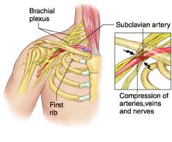
thoracic outlet syndromeThoracic outlet syndrome is a disorder that occurs when the blood vessels or nerves in the space between the collarbone and first rib (thoracic outlet) become compressed. This can result in any of the following symptoms ; 1. pain in your shoulders and/or neck , 2. numbness / pain / tingling down the arm and / or in a finger / fingers. Common causes include physical trauma, i.e. car crash ; repetitive injuries from job or sports related activities; anatomical defects (such as having an extra rib); poor posture( protruding neck / rounded shoulders); and pregnancy.

You may notice symptoms of thoracic outlet syndrome if your job requires you to do a repetitive movement continuously for long periods, i.e. typing on a computer; working on an assembly line; or lifting things above your head. These repetitive activities tend to tighten up neck and shoulder muscles. Athletes, such as baseball pitchers and swimmers can also develop thoracic outlet syndrome from the years of repetitive movements.

Obesity can put an undue amount of stress on your body structures and joints leading to the development of thoracic outlet syndrome. Carrying around an over sized bag or backpack or having somebody sitting on your shoulders for long periods, say at a concert can compress nerve structures etc. and lead to the condition. You may suffer symptoms during pregnancy as muscles, ligaments, tendons and joints come under strain. This is as a result of the release of the hormone relaxin around week twelve of pregnancy, and also due to the increase in bodyweight.

The two most common types of thoracic outlet syndrome are:

1. Neurogenic (neurological) : This form of thoracic outlet syndrome is characterized by compression of the brachial plexus ( nerves coming from the spinal cord (neck). This network of nerves controls muscle movements and sensations in the shoulder, arm and hand. This is the most common cause of thoracic outlet syndrome.

Signs and symptoms include:
• Numbness or tingling in your arm or fingers.
• Wasting of muscles through which the compressed nerve travels; either in the arm or fingers or both.
• Pain in the neck , shoulder or hand.
• Weakened grip in hand / loss of strength in the arm.

Treatment : Often if neurogenic thoracic outlet syndrome is caught early, deep tissue work on neck, shoulders and arm to loosen out tightened muscles; joint mobilisations / manipulation of the cervical and thoracic spine; electro acupuncture along the affected nerve pathway; postural correction exercises, can usually bring great relief within one or two treatment sessions. If the condition is chronic ( i.e. there a month or months), it may take more time for the irritated nerve to settle. You may need to lose weight if obesity is the cause of your thoracic outlet syndrome.

2.Vascular (venous or arterial): This type occurs when one or more of the veins or arteries are compressed in the area between the collarbone and first rib.
Signs and symptoms include:
• Discoloration of the hand (bluish colour).
• Arm pain and swelling, possibly due to blood clots.
• Blood clots in veins or arteries in the upper area of your body.
• Lack of colour in one or more of your fingers or your entire hand.
• Weakened or no pulse in the affected arm.
• Cold fingers, hands or arms.
• Arm fatigue after activity.
• Numbness or tingling in your fingers.
• Throbbing lump near your collar bone.
• Weakness of arm or neck.

Treatment : For vascular (venous or arterial) thoracic outlet syndrome…you need to consult with your doctor. This condition may require thrombolytic medications, anticoagulant medications or surgery.